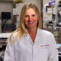
Enjoy the next Thomas Jefferson Point-of-Care Ultrasound Educator of the Month Series! This post is brought to you by Regan Tuder, MD, MS, second-year resident in Emergency Medicine at Thomas Jefferson University Hospital in Philadelphia, PA.
Dr. Henwood sat with the Thomas Jefferson Point-of-Care Ultrasound team to discuss her global health and ultrasound initiatives. She first discussed her interest in global health, which was sparked by a humanitarian medicine trip to Honduras as an EMT and kindled by the 2010 earthquake in Haiti. The earthquake happened to occur during just before a vacation block in her intern year of residency, so she was able to quickly travel to Haiti with a medical field team, learning more about how to practice medicine with extremely limited resources, and integrating ultrasound into her patient care. This experience profoundly changed her practice. She saw firsthand the value in training local providers in ultrasound to continue patient care after the aid organizations left after the immediate crisis. Soon, her focus broadened from humanitarian medicine to ultrasound in resource limited environments. She noted ultrasound was useful for nerve blocks for when pain medicine availability was limited, for triaging patients for transfer, for fractures, distinguishing bladder outlet obstruction from anuria, and making the diagnosis of a hemodynamically-compromising pulmonary embolus. With ultrasound, she saw an avenue to teach a translatable skill that would have real impact on patient care while providing her with multiple areas for research inquiry.
Getting financial support for her non profit organization PURE (Point of- Care Ultrasound in Resource-limited Environments) made the initiation of the organization at times agonizingly slow and occasionally frustrating. Initial funding came through crowd-sourcing and remote mentorship. Trish and her colleagues performed a needs assessment at district hospitals in Rwanda. They evaluated the burden of disease and potential management of patients that could be helped by ultrasound. The ultimate ultrasound education curriculum was vetted by the ministry of health, the medical and dental council, as well as the local university. Rwandan general practitioners were trained partly by remote mentorship and uploading scans through Dropbox for review. The program retains remote mentorships to foster ongoing working relationships and for advice in publishing and writing case reports. The program was quickly expanded to Uganda, and now in several other African countries.
PURE has not functioned without challenges. One continuing struggle is the maintenance skills of practitioners after the initial period of training, a struggle common to ultrasound training programs worldwide. Trish and colleagues have found it is important to have trained on-site teachers at least every few weeks, or at a minimum have one staff member that remains at a program. There is always a challenge in convincing physicians to add new technology to their practice, whether in the US or abroad, and sometimes existing practitioners cannot be convinced to take on a short-term slowdown for long-term improvement in the efficacy of their practice.
In Liberia since the Ebola crisis, there has been funding that allows for a full-time POC ultrasound faculty member and the training of residents in a country that at the start of the training did not have a functioning CT scanner. Prior to this life-saving grant, Dr. Henwood had been watching the response to the Ebola crisis from the beginning, and was one of several physicians who went to Liberia to help. Initially, incorporation of ultrasound was discouraged because of concerns for infection control and the amount of time the examination would take for a practitioner in full protective gear, sweltering on 90 degree days. There were so many types of deaths from the disease, and without a better understanding of the pathology, there was no way to predict which patients had a chance of survival. There was no laboratory testing nor CT available, but ultrasound could distinguish ileus versus perforation in a patient with undifferentiated abdominal pain, or see lung interstitial syndrome (B-lines in lung ultrasound) versus acidosis (none) in a patient with undifferentiated dyspnea. Also, a patient with a fetal demise or missed abortion could be easily determined.
For those teaching in the the USA, Dr. Henwood reminded the group that they may not always be practicing in areas with ultrasound-trained faculty, or CT, MRI, and ultrasound availability in the department of radiology. She urged learners to get used to doing their own scans to feel comfortable in settings with fewer resources. For those with an interest in global health but training in the US, know that the scans in resource-limited areas will have a much higher (about 70%) percentage of some type of pathology. Because of that, there are some additional scans to learn. Physicians should become more familiar with scanning in the 2nd and 3rd trimester, as patients are often presenting later to care. They will likely also see more hepatic abscesses and signs of tuberculosis, and valvular pathology, often related to rheumatic heart disease. For TB specifically, the FASH exam was developed (focused assessment using ultrasound for HIV-associated tuberculosis). It is similar views to a FAST exam with the addition of evaluation for periaortic lymphadenopathy. The exam utilizes windows of six planes to evaluate for evidence of pleural or pericardial effusion, ascites, abdominal lymphadenopathy, and focal splenic or liver lesion or microabscess. Dr. Henwood reminded the group of ultrasound’s inherent limitations. It can be very helpful in narrowing a differential, but may not give a final diagnosis. Dr. Henwood will continue her work in Boston and abroad in both emergency medicine and with PURE. She has just returned from the African Federation of Emergency Medicine conference in Kigali, Rwanda where she spoke about ultrasound as key for the development of emergency medicine across Africa.
Dr. Henwood is the President & Co-Founder of Point-of-care Ultrasound in Resource-limited Environments (PURE, www.pureultrasound.org), a non-profit organization focused on ultrasound education and research in the developing world with current training efforts in Rwanda, Uganda, Zanzibar, and Liberia. Her research has focused on efficacy of training and creating new ultrasound protocols in limited resource environments. She has published a trial on substitutes for ultrasound gel in resource-limited areas. Dr. Henwood works as an emergency medicine physician in the Division of Emergency Ultrasound and is Assistant Professor of Emergency Medicine at Brigham and Women’s Hospital and Harvard Medical School in Boston. She co-chairs the Global Health Subcommittee for ACEP’s Emergency Ultrasound Section. She is an active triathlete and was raised in Philadelphia with her 5 siblings, and is now kept busy chasing her own 1 year-old daughter Madeline.
Resources:
@pure_updates
